Home

Noros obiecta tiran virus electron microscope sticlă plan de vânzări Poartă

CDC - Diagnosis: Electron Microscopy - Rabies

Picture Perfect: Researchers Gain Clearest Ever Image of Ebola Virus  Protein | Okinawa Institute of Science and Technology Graduate University  OIST
Picture Perfect: Researchers Gain Clearest Ever Image of Ebola Virus Protein | Okinawa Institute of Science and Technology Graduate University OIST
Spy" Virus Eavesdrops on Bacteria, Then Obliterates Them - Scientific  American
Spy" Virus Eavesdrops on Bacteria, Then Obliterates Them - Scientific American

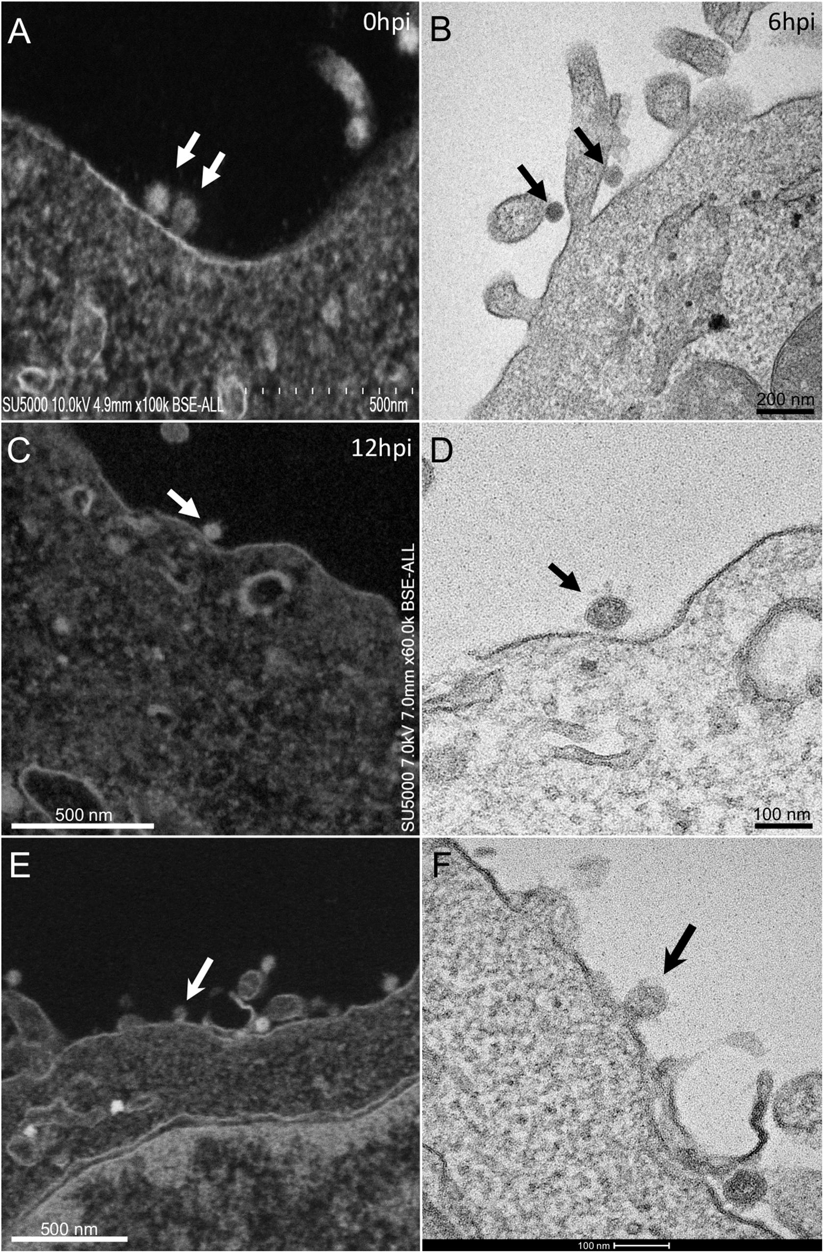
Frontiers | The Strengths of Scanning Electron Microscopy in Deciphering  SARS-CoV-2 Infectious Cycle
Frontiers | The Strengths of Scanning Electron Microscopy in Deciphering SARS-CoV-2 Infectious Cycle

Modern Uses of Electron Microscopy for Detection of Viruses | Clinical  Microbiology Reviews
Modern Uses of Electron Microscopy for Detection of Viruses | Clinical Microbiology Reviews

Viruses | Free Full-Text | Electron Microscopy in Discovery of Novel and  Emerging Viruses from the Collection of the World Reference Center for  Emerging Viruses and Arboviruses (WRCEVA) | HTML
Viruses | Free Full-Text | Electron Microscopy in Discovery of Novel and Emerging Viruses from the Collection of the World Reference Center for Emerging Viruses and Arboviruses (WRCEVA) | HTML
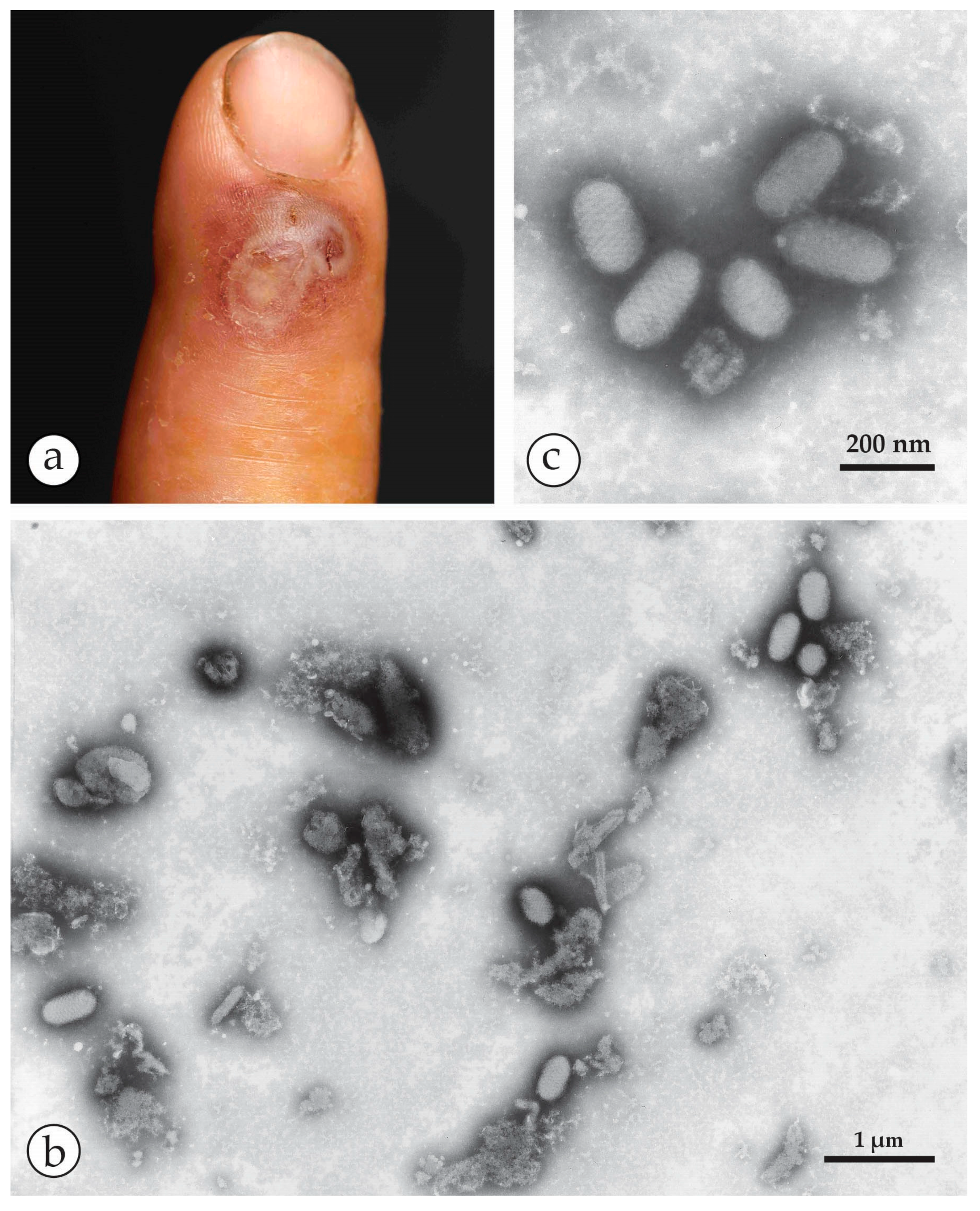
Viruses | Free Full-Text | Rapid Viral Diagnosis of Orthopoxviruses by Electron  Microscopy: Optional or a Must? | HTML
Viruses | Free Full-Text | Rapid Viral Diagnosis of Orthopoxviruses by Electron Microscopy: Optional or a Must? | HTML
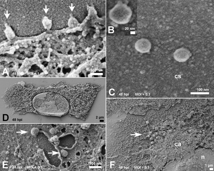
Ultrastructural analysis of SARS-CoV-2 interactions with the host cell via  high resolution scanning electron microscopy | Scientific Reports
Ultrastructural analysis of SARS-CoV-2 interactions with the host cell via high resolution scanning electron microscopy | Scientific Reports

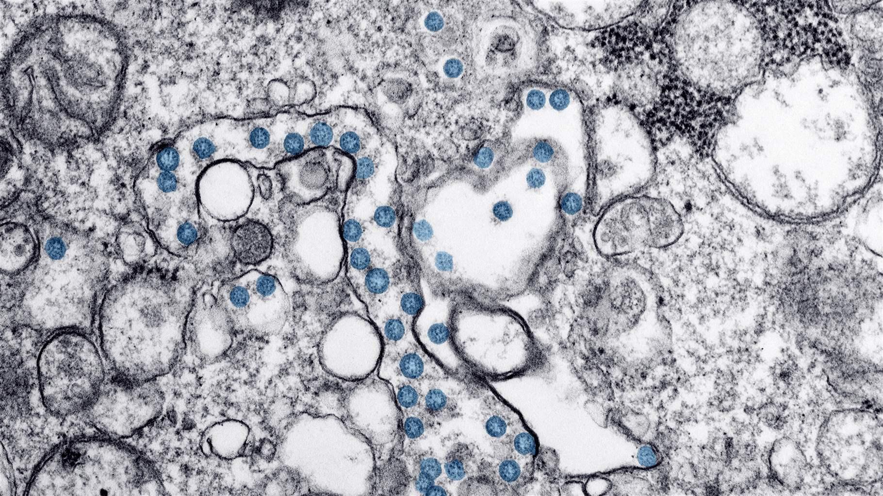
A Look at the Evolution of the Virus That Causes COVID-19 | The Pew  Charitable Trusts
A Look at the Evolution of the Virus That Causes COVID-19 | The Pew Charitable Trusts

Nothing Eats Viruses, Right? Meet Some Hungry Protists - The New York Times

Multimedia Gallery - Transmission electron microscope image shows the virus  in spiny lobster blood cells. | NSF - National Science Foundation
Multimedia Gallery - Transmission electron microscope image shows the virus in spiny lobster blood cells. | NSF - National Science Foundation

Identification of coronavirus particles by electron microscopy requires  demonstration of specific ultrastructural features | European Respiratory  Society
Identification of coronavirus particles by electron microscopy requires demonstration of specific ultrastructural features | European Respiratory Society

Hunting coronavirus by transmission electron microscopy – a guide to  SARS‐CoV‐2‐associated ultrastructural pathology in COVID‐19 tissues -  Hopfer - 2021 - Histopathology - Wiley Online Library
Hunting coronavirus by transmission electron microscopy – a guide to SARS‐CoV‐2‐associated ultrastructural pathology in COVID‐19 tissues - Hopfer - 2021 - Histopathology - Wiley Online Library

Chapter 25 Electron Microscopy of Viruses and Virus-Cell Interactions -  ScienceDirect
Chapter 25 Electron Microscopy of Viruses and Virus-Cell Interactions - ScienceDirect

Viruses | Free Full-Text | Morphologic Differentiation of Viruses beyond  the Family Level | HTML
Viruses | Free Full-Text | Morphologic Differentiation of Viruses beyond the Family Level | HTML

RKI - Consultant Laboratory for Diagnostic Electron Microscopy of  Infectious Pathogens - Vaccinia virus (Poxviruses)
RKI - Consultant Laboratory for Diagnostic Electron Microscopy of Infectious Pathogens - Vaccinia virus (Poxviruses)
Electron Microscopy of Alkhumra Hemorrhagic Fever Virus | Vector-Borne and  Zoonotic Diseases
Electron Microscopy of Alkhumra Hemorrhagic Fever Virus | Vector-Borne and Zoonotic Diseases

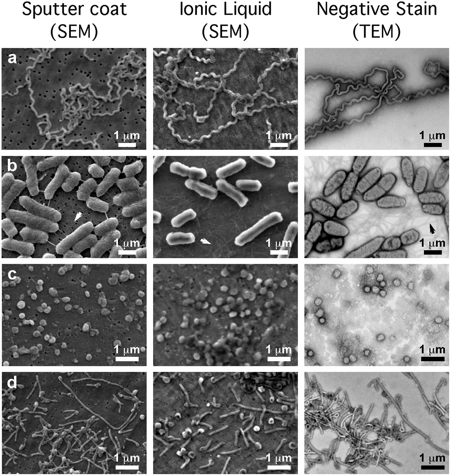
The scanning electron microscope in microbiology and diagnosis of  infectious disease | Scientific Reports
The scanning electron microscope in microbiology and diagnosis of infectious disease | Scientific Reports
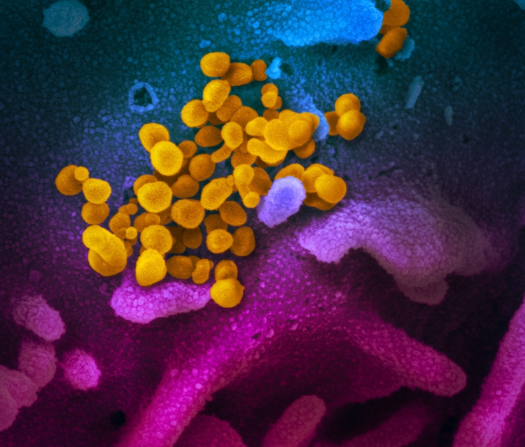
Electron Microscopy of Viruses - ScienceDirect

Image Library | CDC Online Newsroom | CDC

Caution in Identifying Coronaviruses by Electron Microscopy | American  Society of Nephrology
Caution in Identifying Coronaviruses by Electron Microscopy | American Society of Nephrology

New Images of Novel Coronavirus SARS-CoV-2 Now Available | NIH: National  Institute of Allergy and Infectious Diseases
New Images of Novel Coronavirus SARS-CoV-2 Now Available | NIH: National Institute of Allergy and Infectious Diseases

June Almeida discovered coronaviruses decades ago—but got little recognition

Electron Microscopy Images

Ultrastructural analysis of SARS-CoV-2 interactions with the host cell via  high resolution scanning electron microscopy | Scientific Reports
Ultrastructural analysis of SARS-CoV-2 interactions with the host cell via high resolution scanning electron microscopy | Scientific Reports

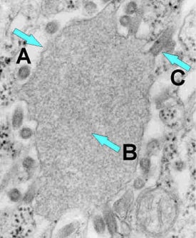
RKI - Consultant Laboratory for Diagnostic Electron Microscopy of  Infectious Pathogens - Monkeypox virus
RKI - Consultant Laboratory for Diagnostic Electron Microscopy of Infectious Pathogens - Monkeypox virus

Modern Uses of Electron Microscopy for Detection of Viruses