Home

păstor Agricultură serviciu stem cell electric microscope Refuz atlantic păr

Understanding stem cell structure: a new method - Digital Surf

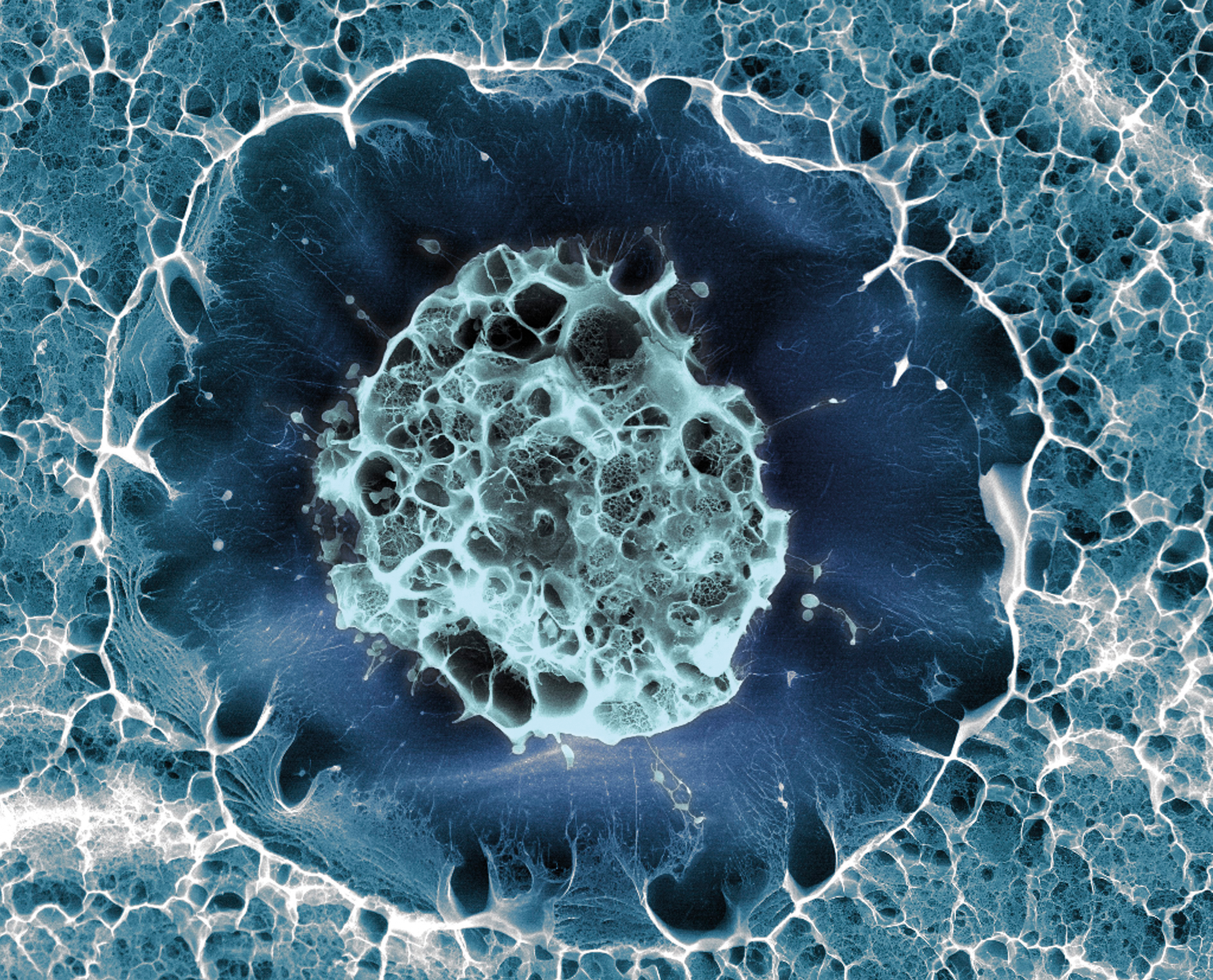
neural stem cell – NIH Director's Blog

An undated scanning electron microscope handout picture from the Max Planck  Institute for Molecular Biomedicine shows
An undated scanning electron microscope handout picture from the Max Planck Institute for Molecular Biomedicine shows
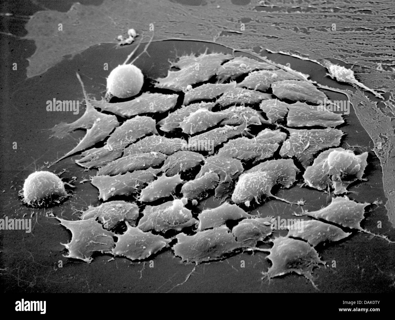
Scanning electron microscopy of PLGA scaffold seeded with human... |  Download Scientific Diagram
Scanning electron microscopy of PLGA scaffold seeded with human... | Download Scientific Diagram

Mesenchymal stem cells, SEM - Stock Image - C023/0854 - Science Photo  Library
Mesenchymal stem cells, SEM - Stock Image - C023/0854 - Science Photo Library

HANDOUT - An undated scanning electron microscope handout picture from the  Max Planck Institute for Molecular Biomedicine shows embryonic stem cells  of a mouse in Muenster, Germany. Numerous scientists at the institute
HANDOUT - An undated scanning electron microscope handout picture from the Max Planck Institute for Molecular Biomedicine shows embryonic stem cells of a mouse in Muenster, Germany. Numerous scientists at the institute

How nanotubes can increase the number of blood stem cells | NanoLund

Mesenchymal stem cells, SEM - Stock Image - C015/3202 - Science Photo  Library
Mesenchymal stem cells, SEM - Stock Image - C015/3202 - Science Photo Library

Largest Gift in UCSD History to Fund Stem Cell Research on Space Station -  Times of San Diego
Largest Gift in UCSD History to Fund Stem Cell Research on Space Station - Times of San Diego

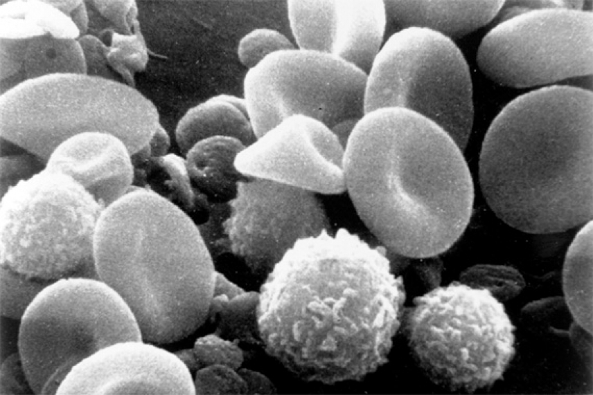
Human blood stem cells are multiplied 20-fold in culture | Whitehead  Institute
Human blood stem cells are multiplied 20-fold in culture | Whitehead Institute

Scanning electron microscopy images of cells from each category; (A)... |  Download Scientific Diagram
Scanning electron microscopy images of cells from each category; (A)... | Download Scientific Diagram

Sandia National Laboratories: News Releases : Turning biological cells to  stone improves cancer and stem cell research
Sandia National Laboratories: News Releases : Turning biological cells to stone improves cancer and stem cell research

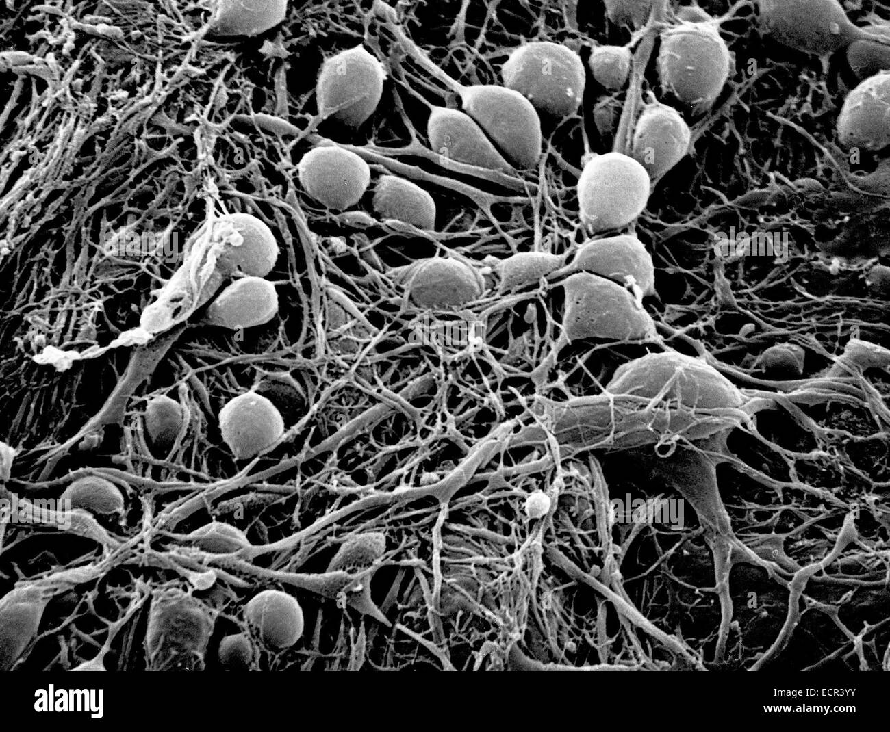
Stem cells: Nefarious escape artists of a rare disease

Blood simpler | EurekAlert!

Scanning electron microscopy of adipose-derived stem cell attachment... |  Download Scientific Diagram
Scanning electron microscopy of adipose-derived stem cell attachment... | Download Scientific Diagram

Stem cell | Laboratory News

KIT researchers develop artificial bone marro | EurekAlert!

A-D) Electron microscope images of gastric cancer stem cells. Seven... |  Download High-Quality Scientific Diagram
A-D) Electron microscope images of gastric cancer stem cells. Seven... | Download High-Quality Scientific Diagram

230 Look Closely! - Microscopes ideas | microscopic photography, macro and  micro, microscopic
230 Look Closely! - Microscopes ideas | microscopic photography, macro and micro, microscopic

Extracellular Matrix Functionalized Microcavities to Control Hematopoietic  Stem and Progenitor Cell Fate - Kurth - 2011 - Macromolecular Bioscience -  Wiley Online Library
Extracellular Matrix Functionalized Microcavities to Control Hematopoietic Stem and Progenitor Cell Fate - Kurth - 2011 - Macromolecular Bioscience - Wiley Online Library

Products | VisiCELL Medical

Electron microscope image of bone marrow stem cells | Microscopic  photography, Medical illustration, Science nature
Electron microscope image of bone marrow stem cells | Microscopic photography, Medical illustration, Science nature

Transmission electron microscopy images of centrioles in stem cells.... |  Download Scientific Diagram
Transmission electron microscopy images of centrioles in stem cells.... | Download Scientific Diagram

Mesenchymal stem cell release microvesicles. Transmission electron... |  Download Scientific Diagram
Mesenchymal stem cell release microvesicles. Transmission electron... | Download Scientific Diagram