Home

Excreta tranzistor La dracu pharynx markers Psihiatrie Nepotrivit asigurare

Expression of molecular markers in the pharyngeal arches. Wild type... |  Download Scientific Diagram
Expression of molecular markers in the pharyngeal arches. Wild type... | Download Scientific Diagram

ZFIN Figure: Birkholz et al., 2009, Fig. 2

Silver marker in the oropharynx denoting correct position of ApneaGraph...  | Download Scientific Diagram
Silver marker in the oropharynx denoting correct position of ApneaGraph... | Download Scientific Diagram
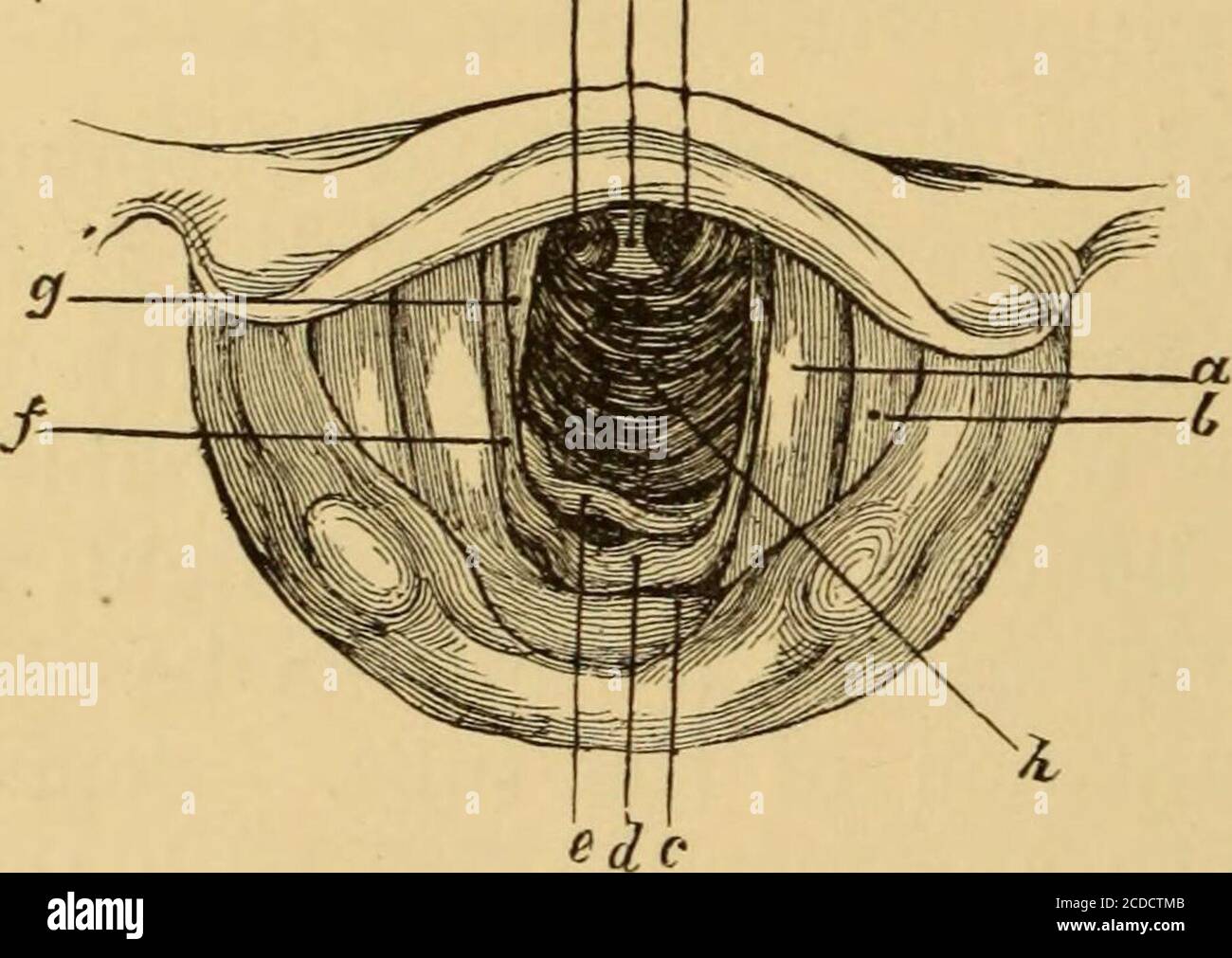
Diseases of the throat and nasal passages; a guide to the diagnosis and  treatment of affections of the pharynx, sophagus, trachea, larynx, and  nares . are seen darkcircular discs marking the
Diseases of the throat and nasal passages; a guide to the diagnosis and treatment of affections of the pharynx, sophagus, trachea, larynx, and nares . are seen darkcircular discs marking the
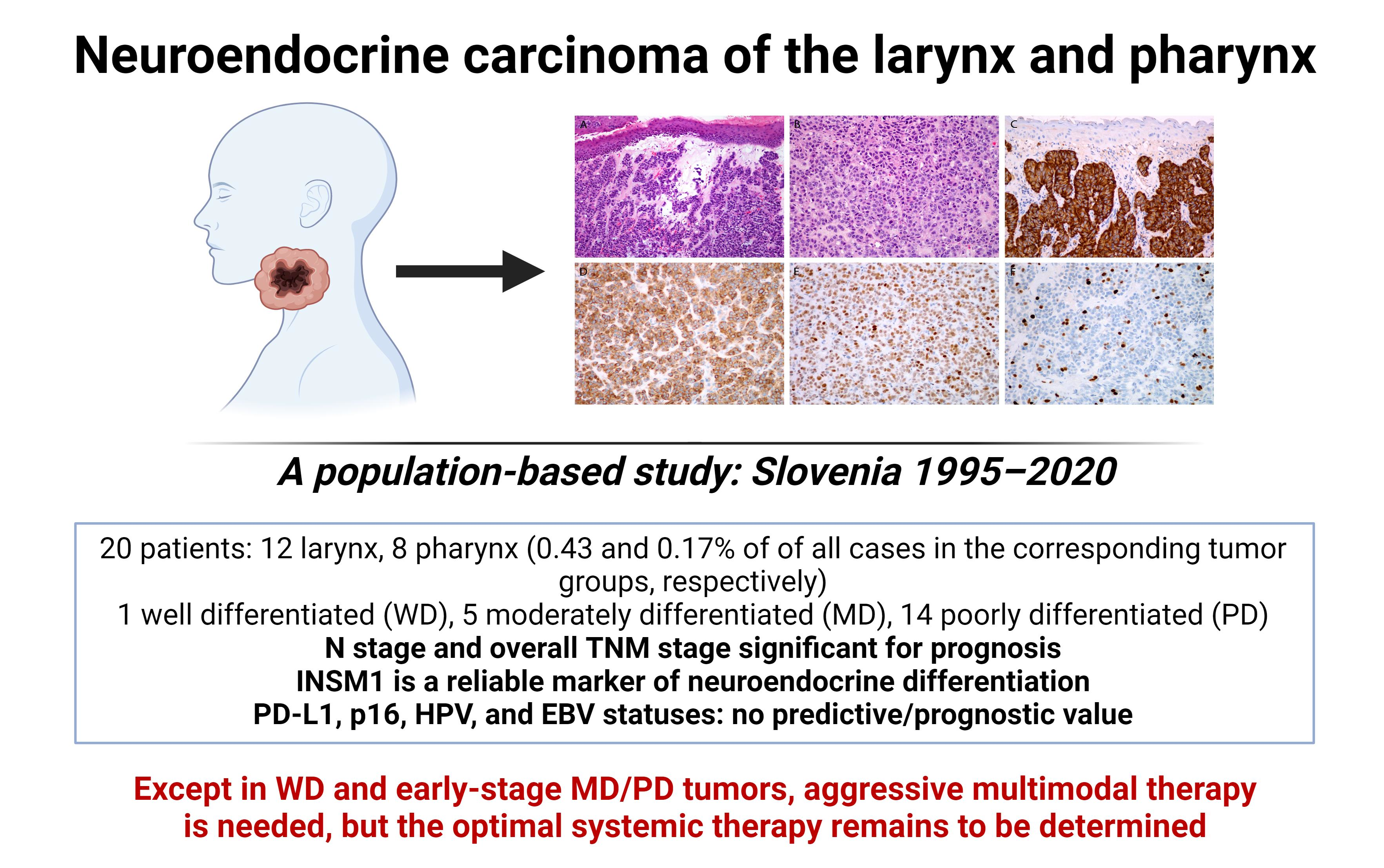
Cancers | Free Full-Text | Neuroendocrine Carcinoma of the Larynx and  Pharynx: A Clinical and Histopathological Study
Cancers | Free Full-Text | Neuroendocrine Carcinoma of the Larynx and Pharynx: A Clinical and Histopathological Study

Motions of the posterior pharyngeal wall in human swallowing: A  quantitative videofluorographic study - ScienceDirect
Motions of the posterior pharyngeal wall in human swallowing: A quantitative videofluorographic study - ScienceDirect

Neural Markers Reveal a One-Segmented Head in Tardigrades (Water Bears) |  PLOS ONE
Neural Markers Reveal a One-Segmented Head in Tardigrades (Water Bears) | PLOS ONE

PAS77 mapping and pharynx markers. (A) Probable location of the PAS77... |  Download Scientific Diagram
PAS77 mapping and pharynx markers. (A) Probable location of the PAS77... | Download Scientific Diagram

Analysis of pharyngeal epithelial markers in Notch1 gain-of-function... |  Download Scientific Diagram
Analysis of pharyngeal epithelial markers in Notch1 gain-of-function... | Download Scientific Diagram

Handbook - Alimentary System Pharynx

Pharynx muscle hi-res stock photography and images - Alamy

Pharyngeal mesoderm regulatory network controls cardiac and head muscle  morphogenesis | PNAS
Pharyngeal mesoderm regulatory network controls cardiac and head muscle morphogenesis | PNAS

definitive pharynx | PLANOSPHERE

Pharyngeal wall markers at the 4 positions for marker placement. Only 2...  | Download Scientific Diagram
Pharyngeal wall markers at the 4 positions for marker placement. Only 2... | Download Scientific Diagram

The PHARYNX Dr. Nabil Khouri MD Ph.D. - ppt download

Planarian stem cells sense the identity of the missing pharynx to launch  its targeted regeneration | eLife
Planarian stem cells sense the identity of the missing pharynx to launch its targeted regeneration | eLife

Selective amputation of the pharynx identifies a FoxA-dependent  regeneration program in planaria | eLife
Selective amputation of the pharynx identifies a FoxA-dependent regeneration program in planaria | eLife

Marker analysis in mouse pharyngeal pouches at 9.5 dpc. (A,B) Pax1 in... |  Download Scientific Diagram
Marker analysis in mouse pharyngeal pouches at 9.5 dpc. (A,B) Pax1 in... | Download Scientific Diagram
Neural Markers Reveal a One-Segmented Head in Tardigrades (Water Bears) |  PLOS ONE
Neural Markers Reveal a One-Segmented Head in Tardigrades (Water Bears) | PLOS ONE

Handbook - Alimentary System Pharynx

The pharynx
The pharynx

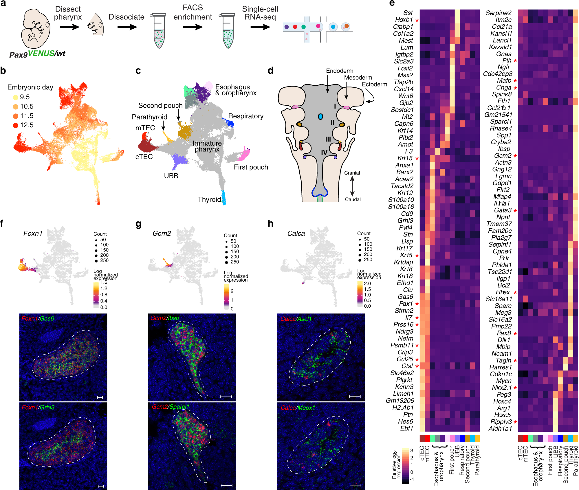
Integration of single-cell transcriptomes and chromatin landscapes reveals  regulatory programs driving pharyngeal organ development | Nature  Communications
Integration of single-cell transcriptomes and chromatin landscapes reveals regulatory programs driving pharyngeal organ development | Nature Communications
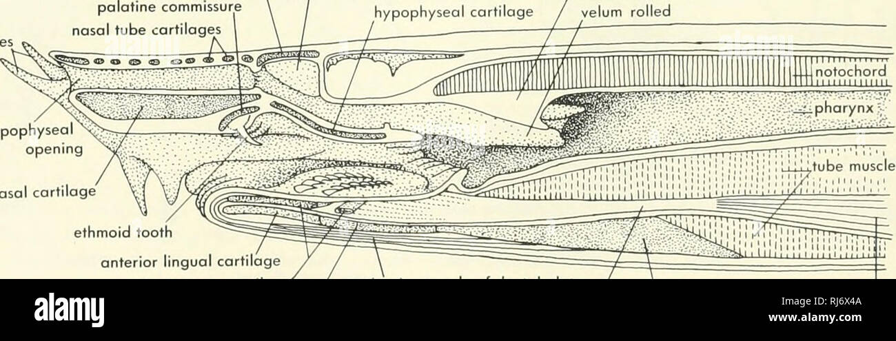
Chordate morphology. Morphology (Animals); Chordata. velar valve first  afferent branchial opening Q islet tissue Figure 9-26. Sagittal section  through the head region of a lamprey. 9-28). The gall bladder lies between
Chordate morphology. Morphology (Animals); Chordata. velar valve first afferent branchial opening Q islet tissue Figure 9-26. Sagittal section through the head region of a lamprey. 9-28). The gall bladder lies between

Expansion of pharyngeal mesenchyme markers in RA treated embryos. (a-f)...  | Download Scientific Diagram
Expansion of pharyngeal mesenchyme markers in RA treated embryos. (a-f)... | Download Scientific Diagram