Home

amăgire puşcă De ce iris cells microscope sexual zahăr categoric

10 Biothechnology Stock Video Footage - 4K and HD Video Clips | Shutterstock

Anatomical structure of the stem of Iris (Juno) magnifica: (a) scheme;... |  Download Scientific Diagram
Anatomical structure of the stem of Iris (Juno) magnifica: (a) scheme;... | Download Scientific Diagram
SEM of iris epithelial cells of the eye - Stock Image - P424/0119 - Science  Photo Library
SEM of iris epithelial cells of the eye - Stock Image - P424/0119 - Science Photo Library

Iris of the eye, SEM - Stock Image - P424/0106 - Science Photo Library

Stomatal Conductance: Functions, Measurement, and Applications - CID  Bio-Science
Stomatal Conductance: Functions, Measurement, and Applications - CID Bio-Science

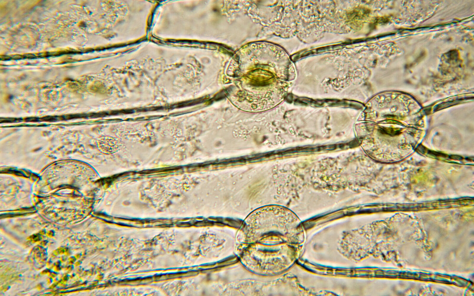
See the Plant Kingdom's Hidden Microscopic Wonders

277 Iris Eye Microscope Stock Photos - Free & Royalty-Free Stock Photos  from Dreamstime
277 Iris Eye Microscope Stock Photos - Free & Royalty-Free Stock Photos from Dreamstime

Living Plant Cells Breathing Stoma Iris Stock Footage Video (100%  Royalty-free) 11783708 | Shutterstock
Living Plant Cells Breathing Stoma Iris Stock Footage Video (100% Royalty-free) 11783708 | Shutterstock

Pigment organelles of pigment cells on the anterior surface of the iris...  | Download Scientific Diagram
Pigment organelles of pigment cells on the anterior surface of the iris... | Download Scientific Diagram

277 Iris Eye Microscope Stock Photos - Free & Royalty-Free Stock Photos  from Dreamstime
277 Iris Eye Microscope Stock Photos - Free & Royalty-Free Stock Photos from Dreamstime

Iris imaging by Scanning Electron Microscopy shows the iris morphology... |  Download Scientific Diagram
Iris imaging by Scanning Electron Microscopy shows the iris morphology... | Download Scientific Diagram

File:Iris germanica root transverse incision with labels.jpg - Wikimedia  Commons
File:Iris germanica root transverse incision with labels.jpg - Wikimedia Commons

Retinal Microscopy
Retinal Microscopy

Confocal microscopy of tissue microarrays – in depth analysis of the  cellular microenvironment.
Confocal microscopy of tissue microarrays – in depth analysis of the cellular microenvironment.

pigment cells Dr.Jastrow's electron microscopic atlas

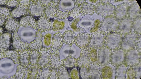
Light micrograph of an iris petal - Stock Image - B060/0040 - Science Photo  Library
Light micrograph of an iris petal - Stock Image - B060/0040 - Science Photo Library

Iris root, light micrograph - Stock Image C002/7006 | Microscopic  photography, Microscopic cells, Micro photography
Iris root, light micrograph - Stock Image C002/7006 | Microscopic photography, Microscopic cells, Micro photography

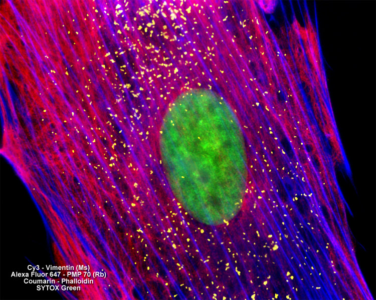
ZEISS Microscopy Online Campus | Fluorescence Image Gallery | Adherent Cells

Live Blood Analysis Microscopes — Microscopes.com.au

How Beautiful Hearing Loss Research Can Be — Hearing Health Foundation

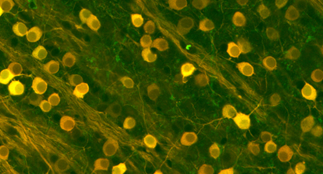
Map of Mouse Iris Offers New Look at the Eye | HHMI

Basic Histology of the Eye and Accessory Structures - EyeWiki

Iris pigment cells, TEM - Stock Image - C049/7149 - Science Photo Library