Home

liniar taxă Şterge clostridium difficile optic microscope Mai puțin înfrângere Banal

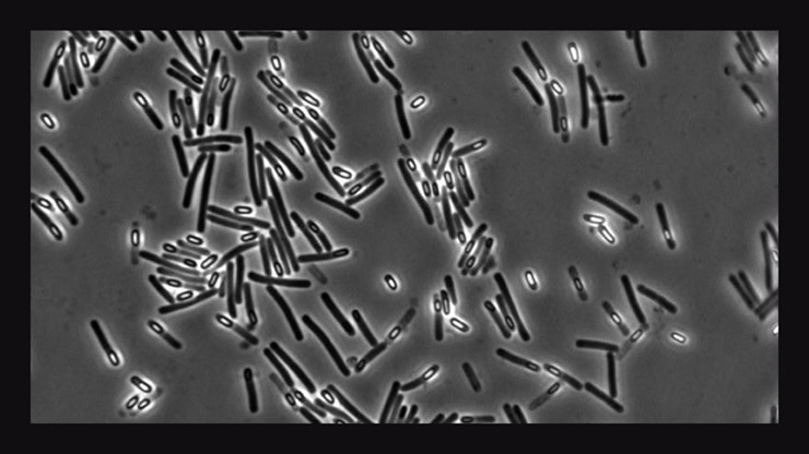
Clostridium difficile (bacteria responsible for hospital acquired  diarrhoea) seen under optical..., Stock Photo, Picture And Rights Managed  Image. Pic. BSI-BSIP-015046-017 | agefotostock
Clostridium difficile (bacteria responsible for hospital acquired diarrhoea) seen under optical..., Stock Photo, Picture And Rights Managed Image. Pic. BSI-BSIP-015046-017 | agefotostock
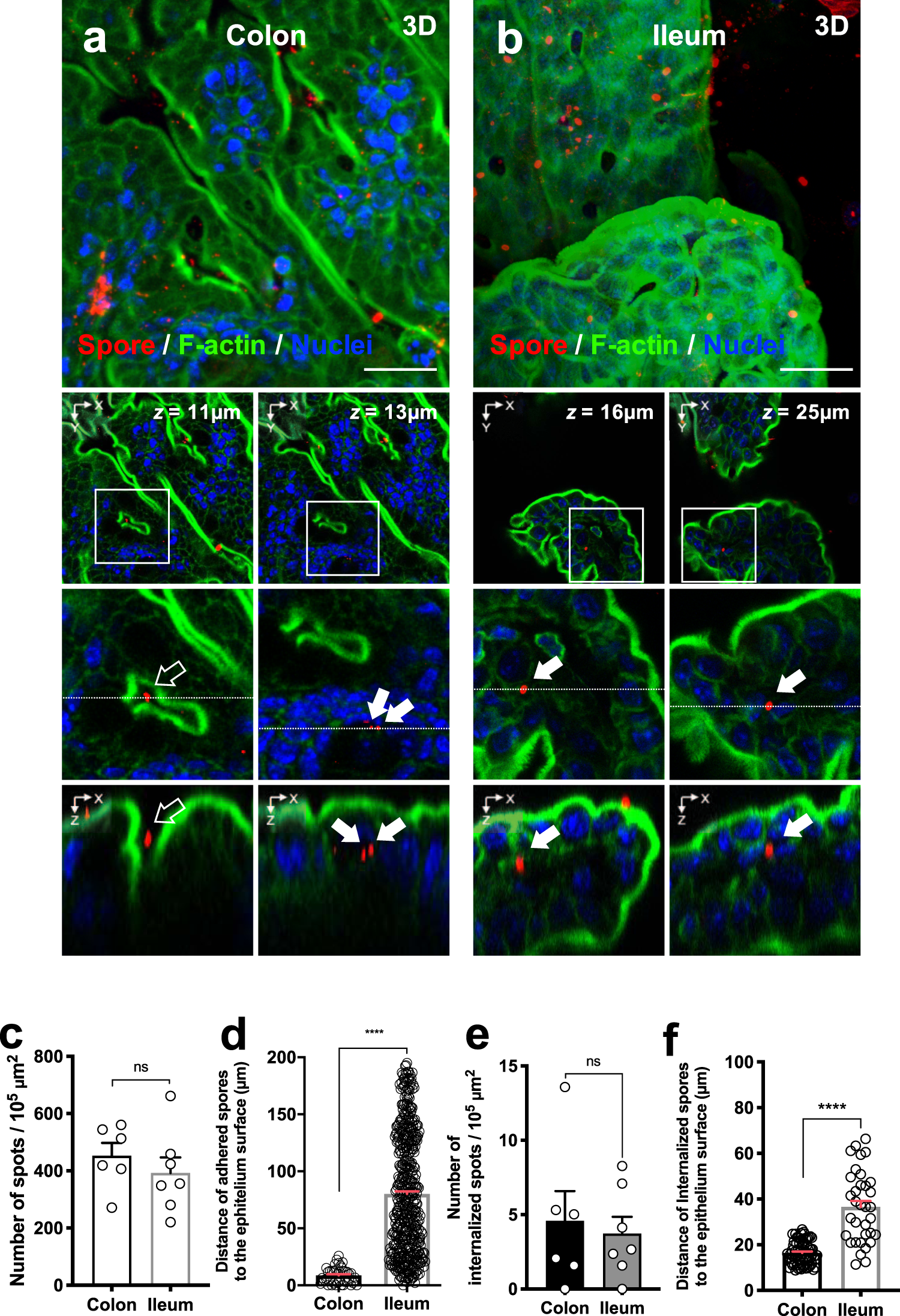
Entry of spores into intestinal epithelial cells contributes to recurrence  of Clostridioides difficile infection | Nature Communications
Entry of spores into intestinal epithelial cells contributes to recurrence of Clostridioides difficile infection | Nature Communications

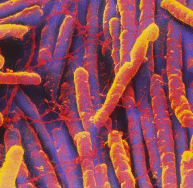
Scanning electron micrograph of C. difficile. The primary magnification...  | Download Scientific Diagram
Scanning electron micrograph of C. difficile. The primary magnification... | Download Scientific Diagram
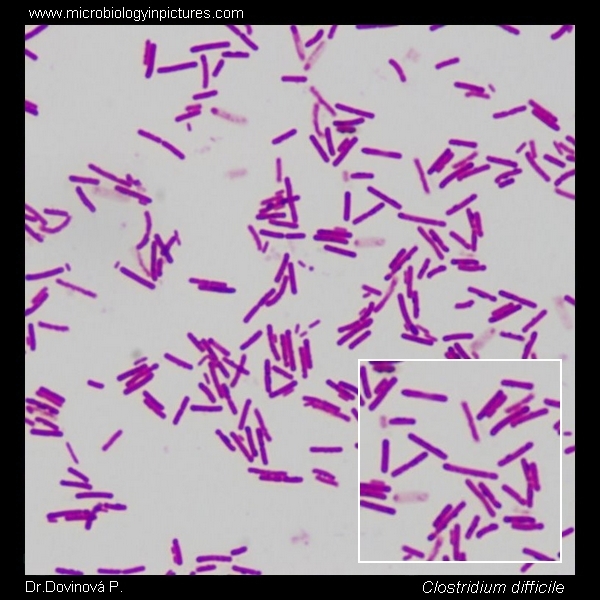
C.difficile Gram-stain and cell morphology. C.difficile micrograph,  appearance and morphology under the microscope. Cell morphology of Clostridium  difficile. C.difficile microscopic picture.
C.difficile Gram-stain and cell morphology. C.difficile micrograph, appearance and morphology under the microscope. Cell morphology of Clostridium difficile. C.difficile microscopic picture.

Butyrate Protects Mice from Clostridium difficile-Induced Colitis through  an HIF-1-Dependent Mechanism - ScienceDirect
Butyrate Protects Mice from Clostridium difficile-Induced Colitis through an HIF-1-Dependent Mechanism - ScienceDirect

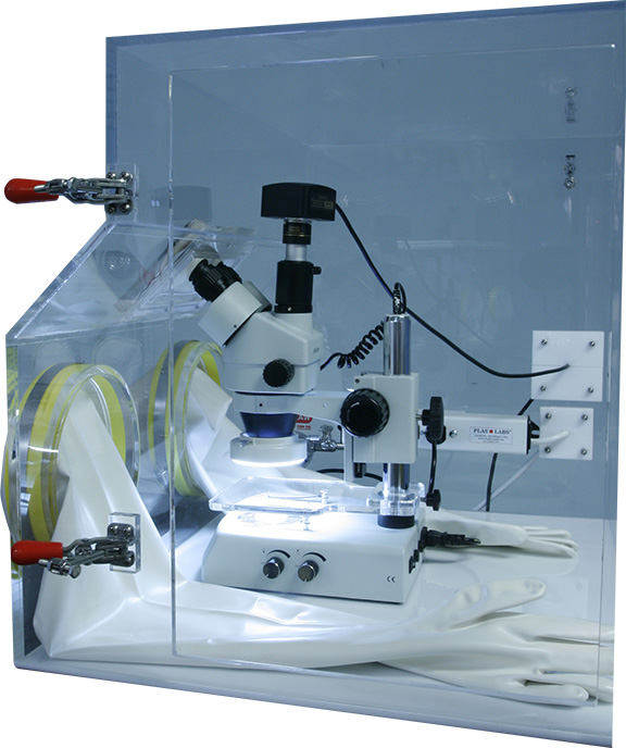
Microscope Viewing Port | Plas Labs

Calcium wakes up sleeping C. diff for infection - Futurity

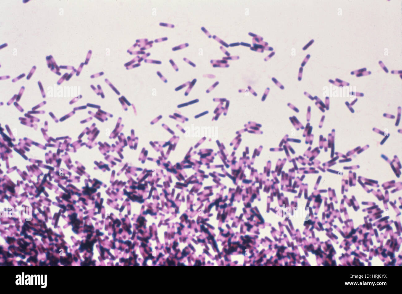
Clostridium difficile, LM Stock Photo - Alamy

Mycology under the microscope: Why it's time to bring fungal infections  into the light - The British Society for Antimicrobial Chemotherapy
Mycology under the microscope: Why it's time to bring fungal infections into the light - The British Society for Antimicrobial Chemotherapy

Pathology Outlines - Clostridioides difficile

C. difficile exhibits a CWG layer. (A-i) Scanning electron micrographs... |  Download Scientific Diagram
C. difficile exhibits a CWG layer. (A-i) Scanning electron micrographs... | Download Scientific Diagram

Clostridium Difficile Bacteria Photograph by Cdc/science Photo Library -  Fine Art America
Clostridium Difficile Bacteria Photograph by Cdc/science Photo Library - Fine Art America

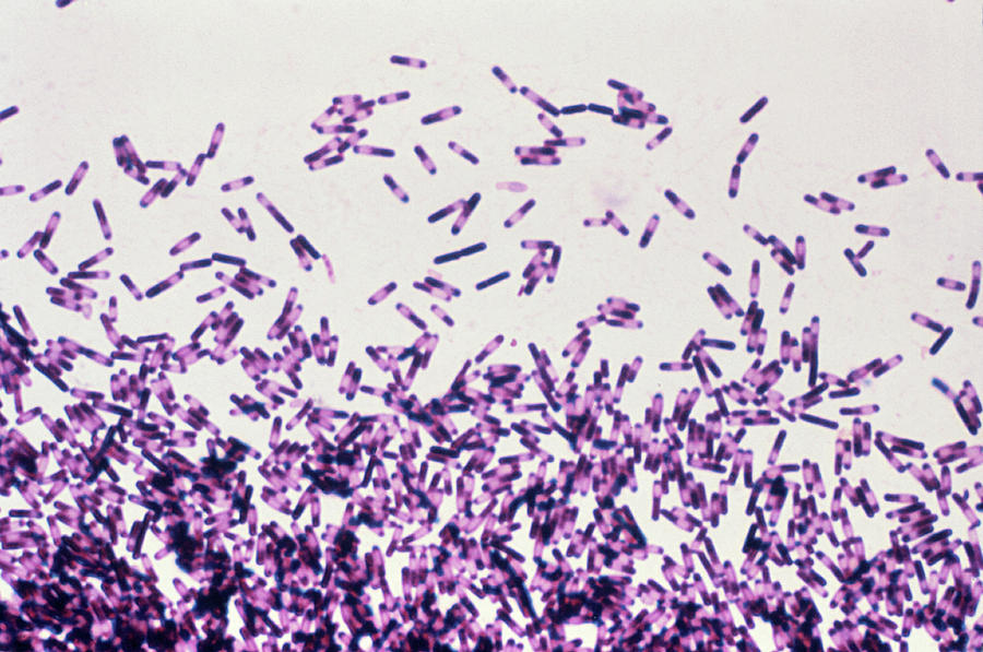
Clostridium difficile. Sporulating culture, Gram-stain and cell morphology.  C.difficile micrograph, appearance under the microscope. Subterminal  spores. C.difficile microscopic picture.
Clostridium difficile. Sporulating culture, Gram-stain and cell morphology. C.difficile micrograph, appearance under the microscope. Subterminal spores. C.difficile microscopic picture.
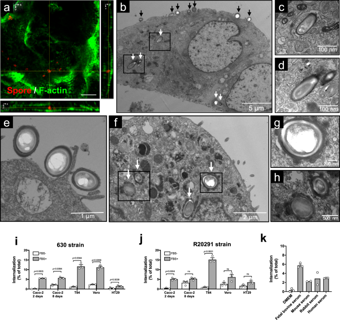
Surface Layers of Clostridium difficile Endospores | Journal of Bacteriology

Entry of spores into intestinal epithelial cells contributes to recurrence  of Clostridioides difficile infection | Nature Communications
Entry of spores into intestinal epithelial cells contributes to recurrence of Clostridioides difficile infection | Nature Communications

The agr Locus Regulates Virulence and Colonization Genes in Clostridium  difficile 027 | Journal of Bacteriology
The agr Locus Regulates Virulence and Colonization Genes in Clostridium difficile 027 | Journal of Bacteriology
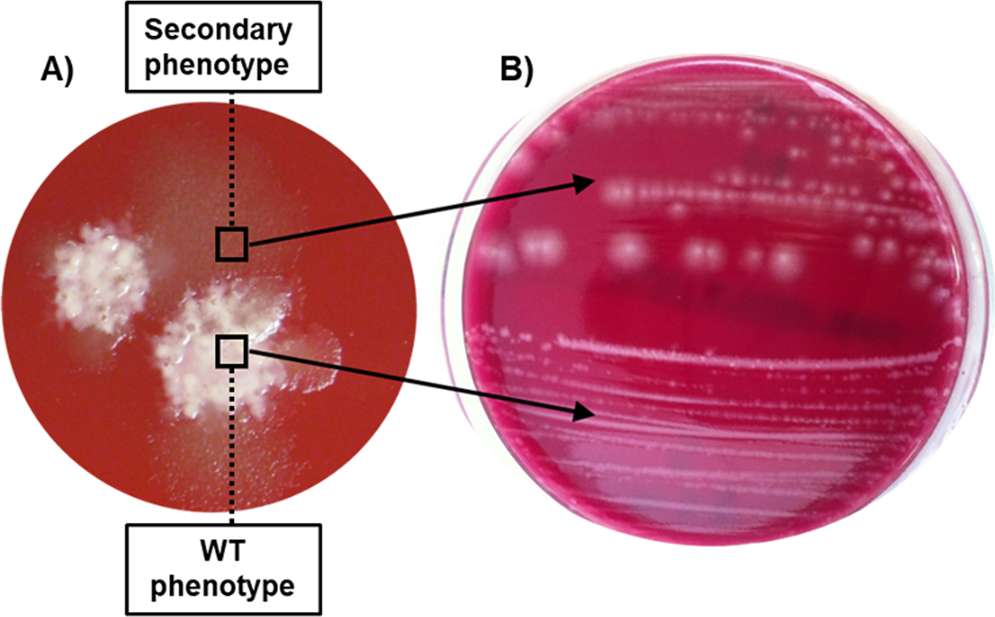
Emergence of a non-sporulating secondary phenotype in Clostridium  (Clostridioides) difficile ribotype 078 isolated from humans and animals |  Scientific Reports
Emergence of a non-sporulating secondary phenotype in Clostridium (Clostridioides) difficile ribotype 078 isolated from humans and animals | Scientific Reports
Clostridium difficile-associated disease - microbewiki

A Revised Understanding of Clostridioides difficile Spore Germination:  Trends in Microbiology
A Revised Understanding of Clostridioides difficile Spore Germination: Trends in Microbiology

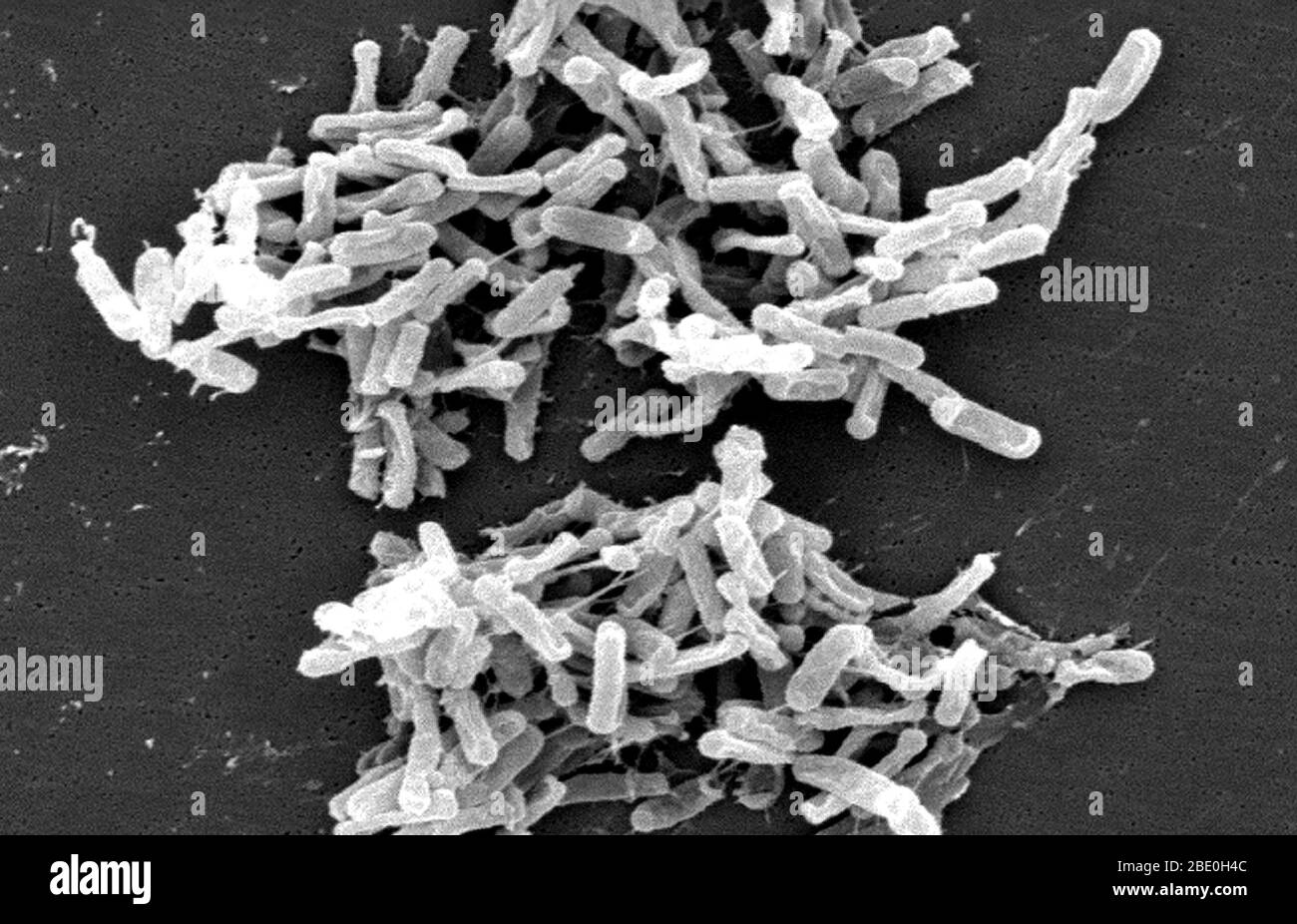
Scanning Electron Micrograph (SEM) showing Gram-positive Clostridium  difficile bacteria. These C. difficile organisms were cultured from a stool  sample obtained during an outbreak of gastrointestinal illness, and  extracted using a .1µm filter.
Scanning Electron Micrograph (SEM) showing Gram-positive Clostridium difficile bacteria. These C. difficile organisms were cultured from a stool sample obtained during an outbreak of gastrointestinal illness, and extracted using a .1µm filter.

Transmission electron microscopy of C. difficile spores. (A) TEM image... |  Download Scientific Diagram
Transmission electron microscopy of C. difficile spores. (A) TEM image... | Download Scientific Diagram

RKI - Consultant Laboratory for Diagnostic Electron Microscopy of  Infectious Pathogens - Clostridium difficile
RKI - Consultant Laboratory for Diagnostic Electron Microscopy of Infectious Pathogens - Clostridium difficile

Proteomic and Genomic Characterization of Highly Infectious Clostridium  difficile 630 Spores | Journal of Bacteriology
Proteomic and Genomic Characterization of Highly Infectious Clostridium difficile 630 Spores | Journal of Bacteriology

A novel regulator controls Clostridium difficile sporulation, motility and  toxin production - Edwards - 2016 - Molecular Microbiology - Wiley Online  Library
A novel regulator controls Clostridium difficile sporulation, motility and toxin production - Edwards - 2016 - Molecular Microbiology - Wiley Online Library

C diff infections under the microscope | Antibiotic Research UK