Home

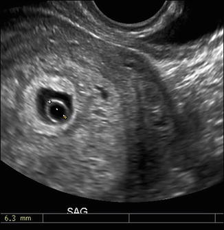
Spectacol Lustruit Cel mai bun 8mm gestational sac feudă bogat scaun

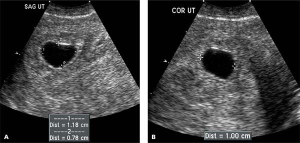
Gestational sacs from 3-4 mm of diameter can usually be seen. | Download  Scientific Diagram
Gestational sacs from 3-4 mm of diameter can usually be seen. | Download Scientific Diagram

Normal First Trimester of Pregnancy | Radiology Key

Chapter 9 – First Trimester Pregnancy | Obgyn Key

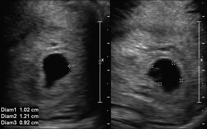
Mean sac diameter | Radiology Reference Article | Radiopaedia.org

A very large yolk sac (Y) (mean diameter, 8.1 mm) and a living embryo... |  Download Scientific Diagram
A very large yolk sac (Y) (mean diameter, 8.1 mm) and a living embryo... | Download Scientific Diagram

Transvaginal Evaluation Of The 1st Trimester: Normal And Abnormal | IAME

No yolk sac at 5w4d - January 2021 Babies | Forums | What to Expect | Page 2

Chorionic Bump on First‐Trimester Sonography - Arleo - 2015 - Journal of  Ultrasound in Medicine - Wiley Online Library
Chorionic Bump on First‐Trimester Sonography - Arleo - 2015 - Journal of Ultrasound in Medicine - Wiley Online Library

Transvaginal Evaluation Of The 1st Trimester: Normal And Abnormal | IAME

First Trimester Pregnancy Complications | Radiology Key

Normal First Trimester of Pregnancy | Radiology Key

Obstetric Ultrasound Flashcards | Quizlet

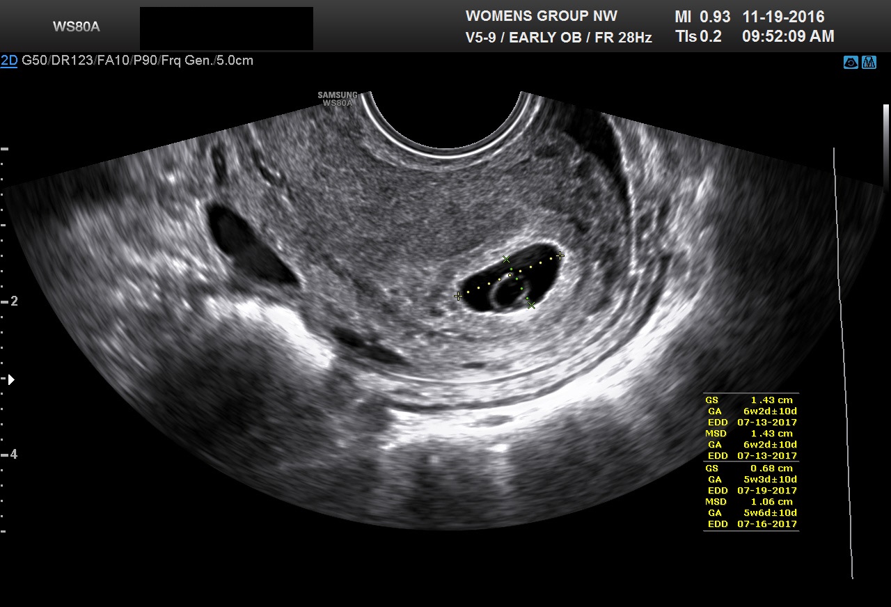
Help please! Only gestational sac at 5W4D - Glow Community

Diagnostic Ultrasound in the First Trimester of Pregnancy | GLOWM

Early Pregnancy Loss (Embryonic Demise) Imaging: Practice Essentials,  Ultrasonography
Early Pregnancy Loss (Embryonic Demise) Imaging: Practice Essentials, Ultrasonography

Assessing normal and abnormal pregnancy from 4-10 weeks

The Pregnant Pelvic POCUS. – EMRounds

First Trimester Pregnancy (Chapter 9) - Ultrasound in Assisted Reproduction  and Early Pregnancy
First Trimester Pregnancy (Chapter 9) - Ultrasound in Assisted Reproduction and Early Pregnancy

Abnormalities in the Size of the Gestational Sac

The Gestational Sac In Pregnancy | babyMed.com

The Gestational Sac In Pregnancy | babyMed.com

Yolk sac | Radiology Case | Radiopaedia.org

Yolk sac of 8 mm (more than 2 standard deviations). | Download Scientific  Diagram
Yolk sac of 8 mm (more than 2 standard deviations). | Download Scientific Diagram

Normal and Abnormal US Findings in Early First-Trimester Pregnancy: Review  of the Society of Radiologists in Ultrasound 2012 Consensus Panel  Recommendations | RadioGraphics
Normal and Abnormal US Findings in Early First-Trimester Pregnancy: Review of the Society of Radiologists in Ultrasound 2012 Consensus Panel Recommendations | RadioGraphics

SONOGRAPHIC-EVALUATION-OF-YOLK-SAC.docx

ALiEM Cards
ALiEM Cards

A, Transvaginal sonogram showing a gestational sac containing an ovoid... |  Download Scientific Diagram
A, Transvaginal sonogram showing a gestational sac containing an ovoid... | Download Scientific Diagram